Techniques
of study Phase 1: Field detection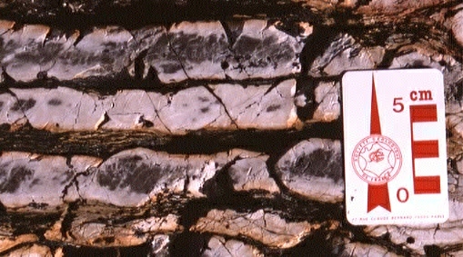 Radiolarians
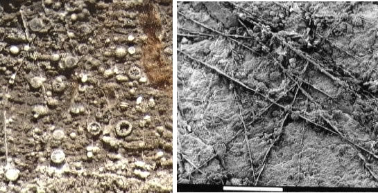
are no bigger than 0.1 - 0.2 mm in average and are not easily detectable
in the field. By applying micropaleontological observation techniques,
the rate of recovery is improved (radiolarian ribbon chert, limestone,
siliceous shale, mudstone, and siltstone). Within suture zones, radiolarian
biostratigraphy is locally the sole tool available for dating siliceous
marine sedimentary rocks. Radiolarians
are no bigger than 0.1 - 0.2 mm in average and are not easily detectable
in the field. By applying micropaleontological observation techniques,
the rate of recovery is improved (radiolarian ribbon chert, limestone,
siliceous shale, mudstone, and siltstone). Within suture zones, radiolarian
biostratigraphy is locally the sole tool available for dating siliceous
marine sedimentary rocks. Close-up of grey radiolarian ribbon chert from the Cache Creek complex, Teslin plateau, Yukon, Canada. |
Phase 2: Radiolarian Chemical Extraction |
|
|
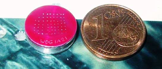 After chemical extraction, the best radiolarian shells are selected and aligned on a small aluminium support (stub) in order to be observed under a scanning electron microscope (SEM). Scale: 1 cent euro coin. After chemical extraction, the best radiolarian shells are selected and aligned on a small aluminium support (stub) in order to be observed under a scanning electron microscope (SEM). Scale: 1 cent euro coin. |
Phase 3: Biochronology and biostratigraphy Radiolarians have a
500 million years age range, making them a powerful biostratigraphic tool.
A selection of adequate and reliable radiolarian assemblages is made through picking techniques. Morphological identification
is performed through optical and/or Scanning Electron microscopes. Illustration: radiolarians
species of Late Devonian, Early Permian, Late Triassic, Middle and Late
Jurassic in age, from left to right. Radiolarians have a
500 million years age range, making them a powerful biostratigraphic tool.
A selection of adequate and reliable radiolarian assemblages is made through picking techniques. Morphological identification
is performed through optical and/or Scanning Electron microscopes. Illustration: radiolarians
species of Late Devonian, Early Permian, Late Triassic, Middle and Late
Jurassic in age, from left to right.
|
 Radiolarian
shells are made of silica (post-mortem recrystallization opal A > opal
CT > quartz). The surrounding matrix of chert, siliceous shale, mudstone,
or siltstone, also contains silica with variable amount of clays. Chemical
processing uses hydrofluoric acid (HF). Techniques related to radiolarian
extraction from siliceous rocks have been discovered at the end of the
1960ies (Hayashi, 1969; Dumitrica, 1970; Pessagno and Newport, 1972). More
recently, these processing techniques have been applied in the field
in relation to geological mapping (Cordey and Krauss, 1990). Left: red radiolarian
chert bed surface after 24 hours of HF processing. Right: detail (scale
1 cm).
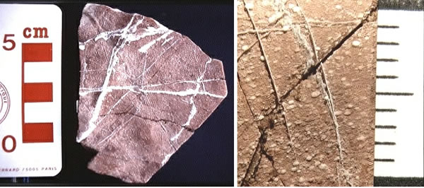
Radiolarian
shells are made of silica (post-mortem recrystallization opal A > opal
CT > quartz). The surrounding matrix of chert, siliceous shale, mudstone,
or siltstone, also contains silica with variable amount of clays. Chemical
processing uses hydrofluoric acid (HF). Techniques related to radiolarian
extraction from siliceous rocks have been discovered at the end of the
1960ies (Hayashi, 1969; Dumitrica, 1970; Pessagno and Newport, 1972). More
recently, these processing techniques have been applied in the field
in relation to geological mapping (Cordey and Krauss, 1990). Left: red radiolarian
chert bed surface after 24 hours of HF processing. Right: detail (scale
1 cm).